Optical Coherence Tomography (OCT)
Smiths Opticians est. 1985
Comprehensive Eye Care with OCT
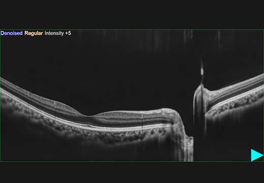
Optical coherence tomography (OCT) is a non-invasive imaging technique used to obtain high-resolution cross-sectional images of biological tissues. It utilizes light waves to capture detailed images of the internal structures of the eye, such as the retina and the optic nerve. OCT is commonly used in ophthalmology to diagnose and manage various eye conditions, including macular degeneration, glaucoma, and diabetic retinopathy. It provides valuable information about the thickness and health of retinal layers, helping eye care professionals to detect and monitor diseases at early stages.
What are the benefits of OCT?
Unless your vision has changed, chances are you don’t give your eye health much thought. But your eyes can actually reveal a lot about your overall health. By looking closely at the retina’s layers, an OCT scan can help detect underlying conditions, including:
- Age-related macular degeneration
- Diabetes
- Glaucoma
- Vitreous detachments
- Macular holes
Lots of eye health problems don’t have visible symptoms until they’re quite advanced, which means that something could be going on behind the scenes without you knowing. By detecting underlying conditions in their early stages, your optometrist can help to manage problems before they get worse.


What happens at an OCT scan?
Short for time? Don’t worry. An OCT scan will be over in a couple of minutes, so there’s no need to put aside any extra time.
Here’s what to expect at your OCT scan:
- First up, a colleague will scan your eyes with the high-tech 3D OCT camera.
- After that, the optometrist will examine the high-resolution 3D images using specialist built-in analysis tools. Once they’re done, they’ll discuss the results with you.
- Next time you come in for an eye test, your optometrist can compare new scan results with old ones. This can help with early diagnosis by detecting the smallest changes to the retina.
